Anatoly. M. Belyaev, Paul V. Yukhalin
Paleovirusology group, Sidose LLC, St. Petersburg, Russia
Email: paleovirusology@mail.ru; abel-7-777@yandex.ru, https://www.paleovirusology.ru/
Despite the diversity of viruses and the role they played in the evolution of the biosphere, their genesis is not clear, since the fossilized remains of viruses have not yet been found in ancient rocks. This is due to the fact that, in essence, viruses were tiny protein capsules that, after death, experienced post-mortem transformations – lysis (dissolution) or collapse.
Keywords: viruses, experimental silification, silification of microorganisms, mineral pseudomorphs, bacterial cell, hot spring.
Experimental silification of bacteriophage T4
The possibility of finding mineral pseudomorphs of ancient viruses is confirmed by the results of experimental silification of bacteriophage T4. Their proteins and icosahedral capsids of the heads were experimentally replaced by silica, which penetrated and deposited in various viral structures (Laidler, et al., 2010). These studies suggest that the silificated microfossils of viruses can be found in rocks.

Experimental silica fossilization of viruses from extremophilic Archaea (rod-shaped SIRV2 – Sulfolobus islandicus viruses, TPV1 – prieurii Thermococcus viruses, and PAV1– Pyrococcus abyssi 1 viruses) confirmed that viruses can be replaced by silica penetrating into various viral structures (proteins, capsids) for several months by analogy with other experimentally and naturally fossilized microorganisms. These studies suggest that viral residues or traces of them may have been preserved in rocks, although their identification may be difficult due to the small size of viral particles (Orange, et al., 2011).
Natural processes of silification of microorganisms
One of the important conditions for preserving the details of the internal structure of microfossils of bacteria, eukaryotes and capsids of viruses is their rapid mineralization, which must occur before the degradation of the bodies of microorganisms. Microorganisms without
a mineral skeleton are best preserved in the processes of silification both near modern thermal springs and in ancient geyserites. Experimental studies of the conditions of silification of cyanobacteria have shown that in heated, silica acid-saturated waters, these processes occur in the interval of several hours, practically during the life of microorganisms (Rozanov et al., 2002). At the same time, microfossils completely retain their volume and are not subject to further digenetic changes (Benning, et al., 2004; Rozanov, et al., 2011).

It is likely that, with the rapid silification of microorganisms, viruses located inside and outside cyanobacteria cells served as centers for the coagulation of silica and the formation of globules.
Silificated globules of virus-like particles inside a bacterial cell were found in the deposits of a hot spring in China (Tucker, 2020).

Fossilization of modern viruses can also occur in special environments called microbial mats, which are found in tidal shoals along coastlines, and in shallow sub littoral lagoons. The substance of microbial mats contains a huge number of viruses – about 30 billion (3×10¹⁰) per gram. At the same time, it was found that viruses mineralize inside the mat due to biochemical processes, photosynthesis and sulfate reduction. Mineralized viruses in the microbial mat have a shape close to spheroidal, but with icosahedral symmetry and form fused clusters (Tucker M., Fossil viruses, 2020).

In laboratory experiments, silificated viruses were also obtained, replaced by iron-containing minerals, iron hydroxyl sulfate (jarosite) and oxides (ferrihydrite and goethite). Such cases have been reported in Rio Tinto (Spain) in the drainage system of acid mines (Tucker, 2020).
Thus, studies of the processes of silification near modern thermal springs, laboratory experiments, studies of modern microbial mats and acid drainage sites show that viruses can be mineralized (fossilized), replaced with calcite, silica and iron minerals. Since lithified microbial mats and ancient geyserites are known in the geological record, ancient viruses can be found in them.
References
- Bacterial Paleontology, Rozanov A.Y., et al., 2002, 188 p.
- Benning L.G., et al., Molecular characterization of cyanobacteria silicification using synchrotron infrared micro-spectroscopy //Geochimica et Cosmochimica Acta. 2004. V. 68. № 4. P. 729–741.
- Laidler, J. R., & Stedman, K. M. Virus silicification under simulated hot spring conditions. Astrobiology, 2010; pp. 569-576.
- Tucker M., Fossil viruses, 2020 // Geology Today· DOI: 10.1111/gto.12321
- Orange, F. et al., Experimental fossilisation of viruses from extremophilic Archaea, Journal: Biogeosciences , 2011 DOI: 10.5194/bg-8-1465-2011
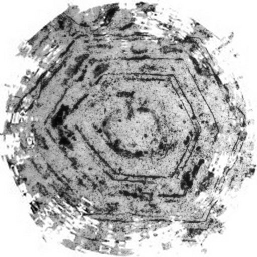
- Zhegallo E.A., et al., Cyanophyta in geyserite constructions of Kamchatka // Algologiya. Vol. 17. No. 1. 2007. pp. 88-92.